


Neurite Bridging Across Micro-Patterned Grooves
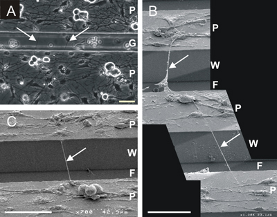
During development and after injury, growing axons must navigate complex, three-dimensional microenvironments. Topographic guidance of neurite outgrowth has been demonstrated in vitro with culture substrates that contain micropatterned features on the nanometer-micron scales. We are characterizing the ability of microfabricated biomaterials to support neurite extension across micropatterned grooves with feature sizes on the order of tens of microns, a size relevant to the design of biomaterials and tissue engineering scaffolds. Neonatal rat dorsal root ganglion (DRG) neurons have been cultured on grooved substrates of poly(dimethyl siloxane) coated with poly-l-lysine and laminin. A subpopulation of DRG neurons displays an unusual capacity to extend neurites that span across the grooves, with no underlying solid support. Multiple parameters influence the formation of bridging neurites and underlying mechanisms are under investigation. These studies are of interest to understanding cytoskeletal dynamics and designing biomaterials for three-dimensional axon guidance.